
Mycobacteriosis
There are several diseases which can affect rainbowfishes. Opportunistic bacteria and parasites can cause dermal and systemic infections. Intestinal nematodes can cause chronic wasting (anorexia) and considerably damage to the intestinal lining. Additionally, water moulds and fungi may also present disease problems in poorly managed aquariums. However, one of the most common and insidious disease of captive rainbowfishes is mycobacteriosis. Mycobacteriosis is a systemic, chronic, progressive disease, and by the time clinical signs or mortality are observed, the disease is already entrenched in the aquarium population.
Two terms are used to describe the disease, either “piscine tuberculosis” or “mycobacteriosis”. The term “piscine tuberculosis” was previously used to describe diseases of fish that involved any acid-fast bacilli. Since the typical tubercular inflammatory response observed in mammals was absent in fish, Parisot & Wood (1960) suggested that “mycobacteriosis” was a more appropriate name for the disease.
Mycobacteriosis is usually a sub-acute to chronic disease of fish where the etiologic agent is an acid-fast bacillus in the genus Mycobacterium. Chronic proliferative mycobacteriosis is characterised by the formation of granulomas, while subacute and acute forms of the disease are associated with necrosis and acid-fast bacilli scattered diffusely among the kidney, liver, spleen, and often all visceral organs. Often no external signs are present until advanced stages of the disease occur, at which time non-specific signs present including emaciation, hemorrhagic and dermal lesions, lethargy, and death. Many Mycobacterium species are ubiquitous in the aquarium hobby and trade, making control by avoidance of these pathogens very difficult. Furthermore, there is currently no effective treatment for mycobacteriosis.
Rainbowfishes appear to be a highly susceptible species based on numerous reports and anecdotal observations. Under pathology examination, mycobacteria have been found in apparently healthy rainbowfishes. The increasing popularity of rainbowfishes has resulted in a significant increase in the number of commercial operators breeding and distributing rainbowfishes. This increases the potential for dissemination and exacerbation of infectious diseases, such as mycobacteriosis. In earlier days you never heard of any rainbowfishes with mycobacteriosis infections. It didn’t start to appear until around the late 1980s. This followed from them being commercially bred and distributed through the aquarium trade. Before then you couldn’t buy rainbowfishes in aquarium stores, you either had to go collect them yourself or get them from other hobbyists. The problem wasn’t so much from the commercial breeders, although I know of one whose breeding establishment was rampant with mycobacteriosis. I believe the problem came from the wholesalers who kept the locally bred rainbowfishes with imported tropicals from Asia and thus eventually becoming infected. Once sold through the aquarium trade and bought by hobbyists the problem became widespread.
Mycobacteria are widespread in Asian fish farms and are commonly found in imported ornamental fish from that source. Asia contributes more than 90% of the world’s ornamental fish production. Like other farming practises, aquaculture is plagued with disease problems resulting from its intensification and commercialisation. Some experts in the aquarium trade believe that mycobacteria infections are becoming more common in imported fish. I believe that it is now so widely circulated in the hobby that it is almost impossible not to be affected by it at some time. It doesn’t really matter how good the wholesalers are in quarantining stock the problem will persist and spread through the fish and holding tanks. Most of the fish won’t show any problems until long after they have been sold to retail stores. Most retails stores will also carry mycobacteria in most if not all of their aquariums. How many aquarium retailers do you know that sterilise their tanks in-between shipments?
This is also the reason I believe that rainbowfishes seem to be more susceptible to mycobacteria than most other aquarium fishes. They have just not had the exposure that the imported fish have had to the disease and therefore have no natural resistance. Rainbowfish breeders who breed from wild stock don’t generally have a problem. However, once the fish are sent to a wholesaler then I would say they have as much chance of catching mycobacteriosis as any other fish distributed through the aquarium trade. Therefore, rainbowfishes should be obtained from specific pathogenfree sources. Knowledge of the origin and aquarium practices of your source can help you prevent potential problems. So the lesson from this is buy your fish direct from well-known reliable breeders or get them from other hobbyists whom you know very well.
Rainbowfishes may also differ from other fish species in their immunologic response to mycobacterial organisms. Eggs of rainbowfishes are released and fertilised externally, and the resulting embryos and larvae are therefore exposed to an aquatic environment full of potential pathogens capable of causing various types of diseases. During the early stages of development, rainbowfish embryos and larvae have little or only limited ability to synthesise immune-relevant molecules endogenously and their lymphoid organs are not yet fully matured. How they survive the pathogenic attacks in such a hostile environment remains relatively unknown. The presence of such pathogens drastically affects reproducibility, fecundity and growth. You need to keep track of the age of your rainbowfishes and avoid breeding fish that are older than 2 years. Breeders should maintain separate brood fish populations.
It is suspected that vertical transmission (transmission from parent to offspring) may occur through egg or sperm products. Vertical transmission of the pathogen allows the continuous and simultaneous spread of the disease from one generation to another. In addition, the obligate intracellular nature of the pathogen enables it to invade cells of other tissues in the vicinity, including the ovary. Thus, ova can be infected either through eggshell contamination from the infected ovarian fluid, or through direct invasion of the pathogen via the egg micropyle. A report from an Australian fish hatchery provided evidence that mycobacteriosis can be introduced by eggs and transmitted to the F1 generation. This observation did not confirm that ovarian transmission takes place, as the egg surface may have been contaminated by peritoneal fluid containing mycobacteria. However, research in 1994 confirmed the transmission of mycobacteria in Siamese fighting fish (Betta splendens), via transovarian passage. Acid-fast bacteria were found in the ova of diseased female Siamese fighting fish, using the fluorochrome technique. Transovarian transmission has also been reported in Danio rerio and Xiphophorus maculatus. The observation of mycobacteria in the piscine ova and tubercle granulomas in the ovary wall suggests that transovarian transmission is a definite possibility.
Clinical Signs
Early signs of mycobacteriosis may be subtle or unapparent, and fulminate clinical signs often do not develop until the disease has become widely systemic. Clinical signs of mycobacteriosis are not specific to the disease and often resemble other diseases. They can vary in occurrence and severity and infected fish may manifest few or no external signs of disease. Clinical signs can vary between fish species and the species of mycobacteria can also influence the clinical symptoms observed. Mycobacteriosis is generally a chronic, slowly progressive disease. The acute form of the disease occurs rarely. It is characterised by rapid morbidity and mortality with few clinical signs. The chronic form of the disease is most commonly seen and it may take months or years for clinical signs to appear. There is ample evidence that these organisms are capable of adapting to prolonged periods of dormancy in tissues, and that this dormancy is responsible for the latency of disease.
Because of the slow progression of the disease, younger fish infected with mycobacteriosis show no external signs. As fish age or are stressed, the infection becomes more serious. It is difficult to specify the length of incubation (the time from infection to the appearance of the first signs of the disease). The incubation period varies greatly and depends on susceptibility, temperature, and severity of exposure.
If clinical signs develop, emaciation, cachexia (wasting, loss of weight), exophthalmia (pop-eye), ascites (dropsy), skeletal deformities (curvature of the spine), haemorrhagic and dermal ulcerative lesions or loss of scales may be observed. Other signs of infection can be seen in the gills, which are paler than normal and show thickened areas on some filaments. Small lesions may be observed around the mouth and vent. Changes in cutaneous pigmentation include a fading of normal colour in aquarium fish or change in colouration. Affected fish generally exhibit lethargic behaviour, isolation, abnormal swimming behaviour, floating impassively on the surface of the water, with concurrent loss of appetite. Poor growth, panophthalmitis and retarded sexual maturation may also occur.
Affected fish populations may show chronic low-level mortality, and increased susceptibility to parasitic infection. Acid-fast staining and mycobacterial culture should be used to evaluate any group of fish showing chronic, low-level mortality and spawning difficulties, regardless of whether they show external signs of the disease. Mycobacteriosis probably predisposed the fish to other pathogens commonly found in the aquarium fishes, such as Aeromonas, Pseudomonas and Flavobacterium.
Source of Infection
Mycobacteriosis disease outbreak in aquarium fish is often reported to be related to management factors. However, even the healthiest aquarium can harbour the bacteria. A variety of bacterial pathogens are always present in an aquarium, even if the system is maintained in optimal condition. Most of them are ubiquitous in aquatic environments and the non-expression of their virulence could be ascribed to a good management of the system and to a good physiological status of the fish. Moreover, the presence of bacteria described as producers of inhibitory compounds, suggests that the indigenous microbiota can control pathogenic organisms in aquarium systems.
Although there is no firm evidence to confirm that environmental stress can cause mycobacteriosis infection, it has been suggested that an unnatural environment, such as an aquarium, may actually promote the disease. Fish should be maintained under optimal conditions. Inappropriate aquarium management can result in abnormal stress and a reduction in the normal resistance of the host. Overcrowding, accumulation of waste and organic matter in the water and increasing water temperature (above 28°C) may all be predisposing factors. Temperature optimums may depend upon the particular strain, but generally range from 25 to 35°C. To a lesser extent, these bacteria also initiate disease in cold-water species. The bacteria can be found in the water column, biofilm and in the top centimetre of the aquarium substrate. They survive in aquarium systems that have a wide range of conductivity, pH and temperature. Attention to water quality and good nutrition will assist the fish in fighting these chronic infections. Poor nutritional health can greatly enhance the progression and severity, and reactivation of disease. The severity of the disease is influenced by a number of interrelated factors, including bacterial virulence, the kind and degree of stress exerted on a population of fish, the physiologic condition of the host, and the degree of resistance inherent within specific populations of fishes. Once present in an aquarium, infection rates can vary from 10 to 100%.
Diseased fish, as well as recovering fishes, may become reservoirs of infection. Thus, contamination from other fish can not be discounted. Mycobacteriosis is thought to be acquired through the ingestion of mycobacteria present in the environment, which usually have their origin in detritus derived from dermal lesions, faecal material, urine or exudates etc., shed by diseased animals that contain mycobacteria. The sources and modes of transmission in fish may be related to the infection of invertebrates, such as freshwater snails, daphnia and shrimp. Live feed for fish, comprising daphnia, mosquito larvae, tubifex, and oligochaetes collected from both the wild and those bred in captivity in the central region of Thailand, were found to be contaminated with M. marinum, M. fortuitum, M. chelonae and other Mycobacterium species. The entry of mycobacteria through skin and gill lesions caused by injury or parasitic infection should also be considered. After the organisms enter the body, they may cause skin lesions or spread to other organs through the circulatory or lymphatic system.
Diagnosis
Unfortunately, there is no non-lethal method available to identify infected individuals, especially those in early to mid stages of disease. Slow mycobacterial growth rates contribute to the late onset and chronic effects of mycobacteriosis. By the time clinical signs and low-level mortality are observed, the disease may already be entrenched in a population. Methods for detection of infected individuals have yet to be developed. The techniques for diagnosing mycobacteriosis in fish are continually evolving, but clinical signs and gross pathology may give an initial indication of infection with mycobacterial species. Most cases of mycobacteriosis in the aquarium hobby are not identified or more often, are simply misdiagnosed. It is recommended that infected fish be submitted to a laboratory for identification. Diagnosis of mycobacteriosis depends on clinical and histological signs and identification of the bacterial pathogen.
Hobbyists Diagnose
The most common bacterial infections in aquarium fish are caused by organisms such as Aeromonas, Pseudomonas, Mycobacterium and Flavobacterium. All these diseases can be spread easily between tanks from contaminated nets, shared equipment, etc. Aeromonas has been found to be the most common. All of them cause opportunistic skin infections often caused by injury or parasitic infection. Mortality increases significantly once bacteria enter the circulatory system. Aeromonas, Pseudomonas and Flavobacterium generally have short incubation period and rapid progression of infection. Clinical signs are generally reached within one week of the initial infection of the disease. On the other hand, mycobacteriosis is a chronic disease and it may take months or years for infected fish to show any clinical signs.
These diseases do not hang around waiting for the average hobbyist to decide what infection the fish have. Aeromonas infections can cause 100% mortality amongst fish in 21 days. The average mortality rate of Pseudomonas can be as high as 50% during the first 21 days with continued mortality for another 7~14 days. Flavobacterium is fast acting and highly contagious. Within 36 hours of infection fish will show areas of greyish discoloration with mortality rates of 30-40% during the first 21 days. Once established, the infection can spread quickly and cause 60-90% mortality rates. Several days can elapse before mortality results from infection by low virulence strains. High virulence strains cause death within 24 to 48 hours post exposure to the pathogen. The infection can be expected to spread most rapidly if water conditions are less than ideal. On the other hand mycobacteriosis is a sub-acute to chronic disease and infected fish populations generally show low-level mortality. Therefore, if you have an infected rainbowfish with a lesion that has not changed that much for more than 21 days, then I would suggest that in all probability it will be a case of mycobacteriosis.
The acute form of the disease occurs rarely. It is characterised by rapid morbidity and mortality with few clinical signs. The chronic form of the disease is most commonly seen and it may take two or more years for the number of organisms to grow to readily detectable numbers. Many species of fish may manifest few or no external signs of disease. Chronic mycobacteriosis infections manifest themselves primarily as swollen white patches or lumps on the body that turn into red or pale lesions. Fish with only skin infections may have several types of concealed lesions. Both the dermis and epidermis are eroded and the underlying musculature becomes severely necrotic. At this stage, the infection has usually become systemic and the infection on the surface of the skin may occur throughout the peritoneum and musculature. Internally the liver, kidney, and spleen may be impaired.
Fish diseases and identification is, at present, difficult. It requires specific laboratory sampling and testing which takes considerable time when bacterial diseases require quick application of treatment. Experienced aquarium hobbyists can and do make accurate, presumptive diagnosis’s based on examination and assessment of the clinical signs, and then apply affirmative control measures. However, economics and other factors will determine the appropriateness of the selected treatment. The cost of treatment may exceed the value of the fish in the aquarium or pond. An aquarist with a large-scale breeding facility stocked with valuable or rare and endangered fish, for example, would probably be wise to spend the money on proper diagnose. On the other hand, if the loss only involved common species, then spending a lot of money for a fish health professional and treatment may not be economically sensible.
Treatment
Immediately remove dead fish and fish that display symptoms of disease or just generally look unhealthy, and keep records of the tanks in which the sick fish were observed. Infected fish can shed pathogens, even when they show no signs of diseases, but especially when they are either morbid or dead. The infected fish should be isolated in a quarantine or treatment tank.
Control of mycobacteriosis in aquarium systems is extremely difficult once an infection has occurred. Currently there are no widely accepted treatments for mycobacteriosis and infections of aquarium fishes should be considered non-treatable. Clinically infected fish or infected populations of fish should be humanely euthanised and immediately destroyed by burning. Unlike most other bacterial fish diseases, there is no cure for mycobacteriosis and it will progress despite your best efforts. The infection will continue, resulting in chronic health problems and eventually, mortality in the whole population.
There are several reasons why systemic mycobacteriosis should not be treated. There is a lack of information on the bioavailability of most chemotherapeutic agents in aquarium fishes, as are successful well-documented clinical trials. In fact, no chemotherapeutic agent is approved for the treatment of mycobacteriosis in aquarium fishes. Efforts to eliminate infection in affected populations with antibiotics have not been successful as mycobacteria are most resistant to conventional antibiotics. Finally, mycobacteriosis has zoonotic potential.
In most situations, the customary treatment for infected fish or populations is euthanasia of the entire stock (especially in breeding facilities), and the disinfection of the aquarium before restocking with clean stock. Fish that have survived an epizootic disease and have recovered may be latent carriers, posing a significant risk to the entire population. It is generally believed that infected fishes are the main source and reservoir of mycobacteria in aquaria. Dead fish, which have died from mycobacteria infection, and live carrier fish, can spread these bacteria. Obviously, the practise of feeding sick rainbowfishes to your pet Saratoga or fluffy feline has its risks. Under no circumstances should fish from an infected population be sold, moved or given away.
Break down the original infected aquarium and any other tank use as a treatment or quarantine tank and disinfect them with a strong chlorine solution. Use Calcium hypochlorite 65% to disinfect any tanks, which are in the vicinity of others housing live fish. Granular chlorine does not volatilise as readily as liquid chlorine (Sodium hypochlorite). In a poorly ventilated fishroom, fumes from liquid chlorine can cause fish kills in adjacent tanks. Concentrations of 200-1000 mg/L available chlorine for 60 minutes should be effective for disinfections of tanks, substrate, and submersed equipment (keep filters running during treatment). In addition, all equipment that has been in contact with the infected fish should be disinfected. Gloves should be worn when handling infected fish or cleaning contaminated tanks or other equipment. Hands should be washed thoroughly afterwards with 70% isopropyl alcohol and a bactericidal soap.
Always use chlorine with caution as repeated use and extended exposure of the silicon sealant to strong chlorine solutions will destroy or render the adhesive bond ineffective on glass aquariums with disastrous results. Chlorine will dissolve synthetic material like sponge filters, but most plastics are unaffected. Calcium hypochlorite is an oxidising agent and should not be exposed to intense heat, acids, or organic compounds because it is a fire hazard, particularly if wet. In some cases, explosion may occur. Always wear eye protection and rubber gloves when handling large quantities of chlorine. Chlorine can be neutralised by adding sodium thiosulfate to the solution (7.5 grams of sodium thiosulfate will neutralise the chlorine present in 5 litres of a solution of 200 mg/L).
However, disinfection is not always successful due in large part to the resistance of many species of mycobacteria to common disinfectants. Mycobacteria are resistant to many commonly used bactericidal agents at standard dosage rates, including chlorine and quaternary ammonium compounds. Mycobacteria can be highly resistant to chlorine disinfection. As much as 10,000 mg/L available chlorine has been reported necessary to kill some species of mycobacteria. Bacterial biofilm in an aquarium can harbour the organism even after aquariums and equipment are disinfected; indeed, biofilm bacteria appear to be more resistant to disinfection than free organisms.
Veterinarians at the National Aquarium in Baltimore, USA recommend using chlorine to clean the tank and substrate, etc., and then spray 65-90% isopropyl alcohol onto the glass, and allow it to dry. They recommend the alcohol as they found that chlorine does not kill all mycobacteria. They use chlorine to remove/oxidise organic material to assure the alcohol contacts all mycobacteria in/on the tank (D. Petty DVM, pers. comm. 1998). Remove all residues of disinfectant from the aquarium before reuse.
Ultimately, the control of mycobacteriosis in aquaria first requires a thorough understanding of the virulence, prevalence, distribution and potential modes of transmission for mycobacteria species found in aquarium systems. There are significant differences in virulence between the various species of mycobacteria that infect aquarium fishes, highlighting the importance of species identification. Preventing the introduction of the highly virulent species or strains to aquarium systems is paramount, because once established, mycobacteria can be very difficult to eliminate. Some species are very common in water and have been isolated from aquaria in the absence of disease (Beran et al. 2006). With these low virulent, ubiquitous strains, dramatic measures such as entire euthanasia of the affected population may not be warranted. However, even with these strains, it would be prudent to minimise the spread of infections. Even in the absence of mortalities, chronic underlying infections are a threat to the overall health of your fishes and it is imperative that disease be minimised.
Antibiotic treatment is usually discouraged due to lengthy treatment times and varying susceptibility of mycobacteria species to different antibiotics (Docostere et al. 2004). However, in circumstances where a stock of fish is very valuable, identification of the mycobacteria species, followed by treatment with the appropriate antibiotic, based on known vulnerability or susceptibility testing, might be advisable upon the recommendation of a qualified fish health professional. Depopulation and disinfection of the aquarium may be necessary when all other measures fail. This unfavourable course of action may be avoidable by minimising the chances of introduction and spread within aquarium facilities.
Clearly, prevention and appropriate routine disinfection should be viewed as the primary means to control mycobacteria in aquarium systems. The quarantine of incoming fish and eggs, regular monitoring, minimising circulating mycobacteria through regular cleaning and maintenance, and upkeep of a UV sterilisation system are the recommended standards.
If more than one aquarium is used, it might be advisable to remove all fish from one system and thoroughly clean and disinfect it from time to time. The chronic nature of mycobacteriosis means that it is often too late for any remedial action to be taken once the first cases have been observed and diagnosed. The same protocol can be used in quarantine systems, at least on a periodic basis, to prevent potential concentration of mycobacteria.
Although fish should be quarantined for at least 4~8 weeks before being placed in their main aquarium, most fish become clinically affected after a longer period of time. Therefore, direct lethal sampling of a quarantined population, with histopathology and culture, may be necessary to detect subclinical infections.
Ideally all equipment such as nets, hoses, buckets, etc. that comes into contact with stock (diseased or healthy) should be regularly immersed into a strong biocide (such as hypochlorite or iodophores), ideally after each use, to achieve sterilisation. At the conclusion of the disinfection process, any residual chlorine can be neutralised by adding crystals of sodium thiosulphate. Any biocide must be rinsed adequately prior to reuse because these compounds are toxic to fish.
Personal Experience
Case # 1: In early 1997, I transferred some 4-year-old Goyder River rainbowfishes from their present aquarium to a larger 600-litre aquarium. I had raised 30 individuals and decided to split them in half. The 600-litre aquarium contained a mixture of fully-grown rainbowfishes. Most of them were more than 4 year old with some specimens as old as 9 years. Over the ensuing weeks the Goyder's, one by one, stopped feeding and started to ‘hang’ just below the surface of the water. Apart from laboured breathing, and looking as though they just had a very large meal, no other symptoms were apparent - death followed soon after. When only 3 individuals were left from the original 15 that had been transferred, I decided that I needed some confirmation of their disorder. I took the remaining three fish to veterinarian, Dr. Stephen Pyecroft BVSc of Aquatic Diagnostic Services International Pty Ltd for examination.
Stephen's diagnose showed that “Disseminated caseating pyogranulomatous inflammation possibly due to Mycobacterium infection” and “Hepatic Lipidosis” (fatty liver disease). He commented, “The severity and chronicity of the pyogranulomatous inflammation suggests this is the primary disease process. Special stains have shown the presence of acid -fast bacilli consistent with Mycobacterium spp. These organisms were found in the macrophages in the liver and kidney. The hepatic lipidosis is quite severe and could well be associated with hepatoencephalopathy although histological evidence of this was not detected in the brain sections examined. The lipidosis was found in all those examined.”
The fish I concentrated on for the histopathological examination definitely showed the greatest degree of pathology and because of the diagnose of mycobacteriosis we must suspect that the total clinical picture observed is due to this problem. There is no ignoring the fact that these fish on the whole were over weight and that the hepatic lipidosis present would have eventually caused their demise had the TB not caused their final problems. The picture is still not that clear and I personally believe that the nutritional imbalance leading to the lipidosis is the major management factor that will need to be addressed. However the fact that a mycobacterial infection is present must, in these fish, be accepted as the primary cause of disease.”
What that means in layman terms is that the fish were overweight and infected with mycobacteria. My conclusion from all this was that the 600-litre aquarium was the culprit and knew somewhere down the track that I would have to destroy all the fish and sterilise the tank with chlorine. This belief was confirmed as I continued to have disease outbreaks in this aquarium with some fish displaying similar symptoms while others also developed external lesions. This aquarium was treated with a strong chlorine treatment and all fish and plants destroyed. It is interesting to note that the remaining 15 fish, two years later and 6-years old, in the original aquarium were still doing well, albeit on a somewhat reduced and modified diet, and showed no external signs of the disease whatever.
Case # 2: About 8 months after the above episode I presented Stephen Pyecroft with six young (1-year-old) specimens of Melanotaenia oktediensis. All six specimens had what I refer to as “Blackhead Disease” in varying degrees. This disease exhibits itself as a black darkening of one side of the head only. Two of specimens also had small skin eruptions on one side of the body and one also showed the darkening skin colouration along one side of the posterior portion of its body. The most severely affected fish would swim with their head up and tail down and showed an increased respiratory rate. This disease (blackhead) seems to be common among rainbowfishes as I have seen it often and many other hobbyists have spoken to me about this problem. It also seems to be particularly prevalent among Goyder River rainbowfish.
Stephen found that most of the fish had enlarged kidneys, which had a granulated pale colour and protruded beyond their normal position. Granulomas were also present in the spleens and around abdominal organs. Acid fast (Ziehl-Nielsen) stains were preformed on impression smears from most of the affected organs and the presence of acid fast bacteria was confirmed. The Diagnose: Disseminated granulomous inflammation - nephritis, hepatitis, and peritonitis.
Stephen comments were “As we have discussed before, the dark areas on the skin are most likely due to a malfunctioning in either the pigment cells or the nerves that control the pigment cells in that area of the skin. The findings of a generalised infection with Mycobacterium species would be suggestive that the localisation of the dark pigmentation is due to the formation of local abscesses, which are then causing the expression of the major clinical sign. Most of these cases of “blackhead syndrome” in rainbowfish that I have investigated have had a primary infection with mycobacteria. There may be other primary causes of this distinct clinical sign but in these fish it was piscine TB.”
Zoonosis
Mycobacteriosis is different from most other fish diseases that you are likely to experience in your aquarium. This is because mycobacteria are capable of causing a wide range of dissimilar symptoms in infected fish and its ability to cause disease in humans. Human infections caused by mycobacteria transmitted from fish or the aquatic environment is quite common. Reports on mycobacterial infection of the skin have been appearing with increasing frequency in the medical literature.
Mycobacteria have a well-documented zoonotic history. In the past, human mycobacterial infections were sporadic and most commonly associated with contaminated swimming pool water (Hellerström 1939; 1951). For this reason, the skin infection was termed swimming pool granuloma. Chlorination practices used today have greatly minimised the frequency of outbreaks from these sources. Since then however, several authors have noted the association of the skin infection with aquariums and today it is generally referred to as “fishkeeper’s disease”. Swift & Cohen (1962) are usually credited with reporting the first case of fishkeeper’s disease. Despite this innocuous nickname, infections often warrant long-term medical treatment and sometimes, even surgery.
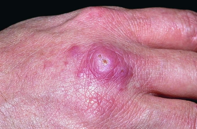
Mycobacteria infection of aquarium hobbyists typically occurs when mycobacteria gains access through skin abrasions and generally produces superficial and selflimiting lesions involving the cooler parts of the body such as hands, forearms, elbows and knees, following direct contact with an infected fish, or contaminated aquatic environments.
Clinical observations typically involve painful or painless reddish nodules at the site of infection, usually adjacent to a cut or scrape. However in several instances lesions developed in an ascending proximal fashion strongly suggesting sporotrichosis. Sporotrichoid skin lesions have a characteristic lymphangitic spread with nodules that ascend proximally along lymphatic vessels. Aquatic mycobacteria can also occasionally spread to the internal body systems of humans and have been isolated from pulmonary lesions, and from synovial fluid and muscle.
Allergic dermatopathies have also been reported on the skin of aquarists handling water in which affected fish have been reared. While these infections are rare, even among those who frequently work in their aquariums, there is at least one case of a child exhibiting multiple lesions spread over the entire body after being bathed in a tub that had recently been used to clean an aquarium. However, “fishkeeper's disease” is not a focal infection of the skin. A case of mycobacteria infection contacted from mouth syphoning water from a fish tank has been reported. It concerned an individual who experienced a throat infection that wouldn't get better, and was eventually diagnosed as fishtank granuloma (Practical Fish Keeping, Jan. 1998). So next time you do a water change and take a big suck on the end of the syphon hose - just think of this article.